3D tomosynthesis can detect up to 35% of cancers sooner.
Since 3D mammograms give a more defined picture, the radiologist can provide a more confident assessment. This means women are less likely to be called back for another scan or have a biopsy.
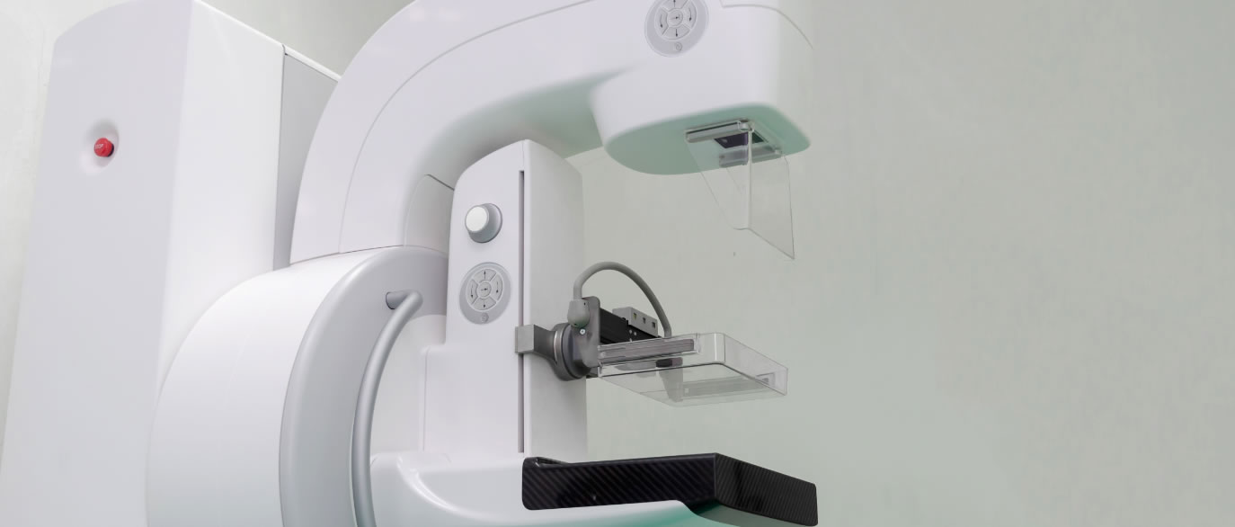
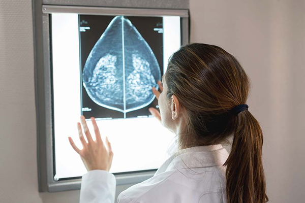
What is a 3D Mammogram?
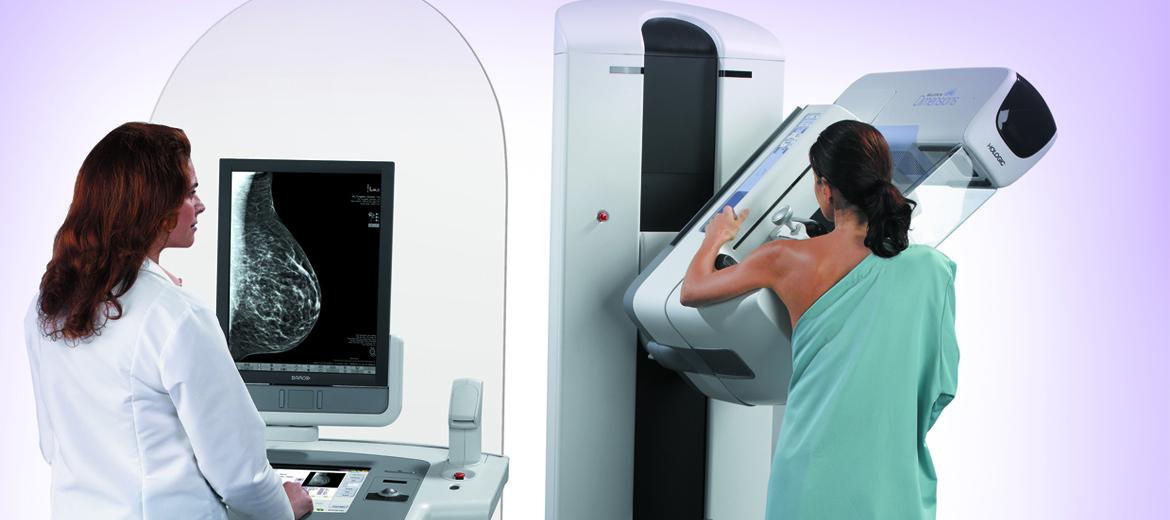
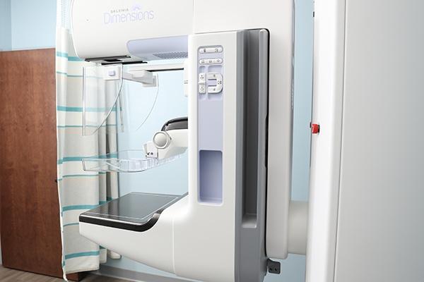
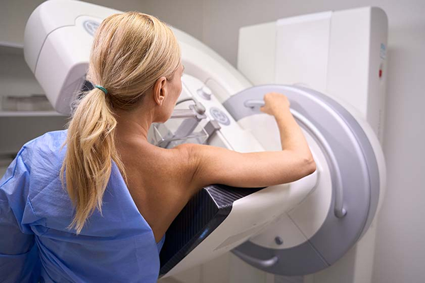
A 3D image of your breast provides better images for screening and diagnosis. It's like seeing the pages inside a book, instead of just the front and back cover. During the exam, the X-Ray arm sweeps in a slight arc over your breast, taking multiple images that create a 3D image of your breast tissue.
What Can I Expect?
The experience of a 3D mammogram is similar to that of a standard mammogram and can be scheduled at an Imaging Center for Women. There is no additional time or additional compression required. Very low X-Ray energy is used during the exam, about the same amount as a traditional mammogram.
How Can I Prepare?
Do not schedule a mammogram the week before or during your period
Do not apply deodorant, perfume, or powder on the day of your mammogram
Wear two-piece garments such as a top and pants or a skirt, rather than a dress