
Diagnostic imaging is the term used to describe all the different ways that physicians are able to look inside your body. A diagnostic imaging center has equipment and trained specialists to perform those tests. A doctor is then able to take the results from the diagnostic tests and draw conclusions from them.
Screening is used by healthy people to look for potential problems. If possible, it tries to detect them even before the patient experiences any symptoms. Diagnostic imaging is used to monitor an existing health condition, investigate a concerning symptom, or pin down a diagnosis. It can be used to guide treatments so that the doctors can give the least invasive treatments possible.
Diagnostic imaging centers use several kinds of imaging machines. They can be used separately or combined depending on the patient’s needs.
Diagnostic Imaging Center Machines
X-Ray is the oldest kind of diagnostic imaging and uses radiation to form images. Solid tissue makes whiter images, so bones show up very white in x-rays. X-rays can be used to examine the skeletal system, to look at objects in the body, and to detect health issues like pneumonia and certain kinds of cancer.
Mammograms use a form of low-dose x-rays to get a clear image of the breast. Doctors are looking for suspicious tissue formations which may indicate breast cancer.
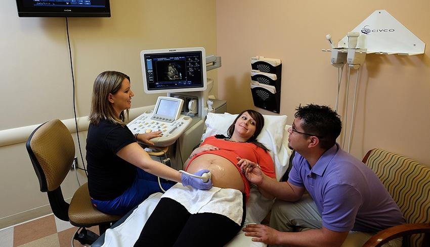
Ultrasounds use high-frequency sound waves to form pictures of what is going on in the body. The most familiar use of this is as a part of prenatal care, but ultrasounds are also used to look for cysts, at your heart and blood vessels, and at your kidneys, liver, and many other organs.
Magnetic Resonance Imaging (MRI) uses a large magnet and radio waves to look at organs and internal structures. It is used to evaluate a wide range of conditions, from tumors to ligaments. It is often used to see the brain and spinal cord.
CT Scans makes cross-sectional pictures of the body using many x-rays taken from different angle and computers to fit them together. It is used to look at the skeleton, to check for blood clots, to look for cancer, internal bleeding, or signs of heart disease. It is most useful when looking at solid tissue.
Positron Emission Tomography (PET) Scans are frequently used in conjunction with CT scans, with CT scans looking at the solid structures and PET scans examining the chemical activity going on in the softer tissues. PET scans use a mildly radioactive drug called a tracer to look at how your tissues are functioning. It is useful in examining cancers, heart disease, and brain disorders.
Fluoroscopy uses x-rays to show a real-time picture of the inner workings of the body. Instead of still slices, it shows actions – the inner workings of swallowing, the actual beats of the heart. It is useful for seeing what is actually going on as it occurs – if you are having trouble swallowing, it will show the exact motions of body structures while swallowing happens. This is very helpful when planning treatments.
Diagnostic imaging centers can use these images to plan and perform minimally invasive procedures as well. The placement of a stent, directing radiation at cancer, and biopsies are all medical treatments that make use of diagnostic imaging.
Need to schedule an appointment? Call us today.


