

Often when people think of MRIs, they think of loud, alarming noises, claustrophobia, and long periods of stillness. But that doesn’t have to be the experience. Now, more hospitals and imaging centers are turning to open MRIs as a more comfortable and stress-free alternative to traditional MRIs. If you or a loved one has recently been prescribed an MRI, it’s important to get the facts before setting up an appointment.
What does “MRI” Stand for?
“Magnetic Resonance Imaging” (MRI) is a tool used to capture images of your body’s soft tissues. MRIs allow physicians to diagnose a wide range of:
- Diseases
- Injuries
- Tumors
- Strokes
- Other issues in the soft tissue that are unreachable by ultrasound or x-ray
What is an MRI used for?
An MRI is able to diagnose a wide variety of injuries and diseases such as:
- Tumors
- Developmental anomalies
- Multiple sclerosis (MS)
- Stroke
- Dementia
- Infection
- Chronic headaches and causes
- Injuries in cartilage or bone
- Herniated discs
- Pinched nerves
- Spinal cord compression
- Fractures
Functional MRIs (fMRI) can also be used to study brain structure and activity, which can help determine neurological issues. MRIs are painless, non-invasive, and radiation free. The machine uses strong magnetic fields and radio waves to capture detailed images on a computer, which a physician then reviews.
How do MRIs work?
Imagine taking a bucket of oil and water and spinning it around. When you stop spinning, the oil will eventually separate from the water, but it might take some time and you can watch it happen. This is kind of like what an MRI does to the atoms in your body.

An MRI combines a powerful magnet with radio waves and a computer to create black and white images. The magnets in the MRI align with the protons in the human body. Radio waves then create a magnetic current, causing the protons to spin away from their previous alignment. When the current is turned off, the protons realign to their magnetic field. Protons in different types of soft tissue take varying time to realign, creating a highly detailed image that can show health conditions and injuries.
A contrast fluid or dye may be injected intravenously to help create clearer images of certain tissues. The contrast fluid makes veins stand out more against organs and soft tissue and can help physicians correctly diagnose issues in complex areas. In any MRI, the patient must remain still in order to achieve the best quality images.
Introducing the True Open MRI
With every new innovation, MRIs become more comfortable. Nowadays, patients can relax when they are told they need to have an MRI. Many larger patients, children, or patients with claustrophobia who previously were unable to get an MRI now have more options available.
A True Open MRI is open on all four sides, providing airflow, a clear line of sight, breathing room, and added comfort for the patient. Many patients who are uncomfortable with enclosed spaces prefer the comfort of the True Open MRI. It can also be a better choice for children who get nervous in a closed MRI.
Open MRI Machines: Dealing with Claustrophobia and Anxiety
For people who suffer from claustrophobia and anxiety, the idea of a traditional MRI could be a bit unpleasant. Wide bore MRIs can offer a bit more space and breathing room, but a True Open MRI can be a complete diagnostic game-changer. Talk to your doctor to see if a True Open MRI is the right fit for your diagnostic needs.
Other types of MRI machines include:
Wide bore MRI: Sometimes referred to as an open MRI, a wide bore MRI opens up the MRI cylinder by an additional 4 inches (70 cm total), which can accommodate larger patients or those with claustrophobia. The image quality is the same as the standard bore MRI, and patients gain a little bit more room to breathe.
3T MRI: A 3T MRI operates at twice the strength of a normal MRI, using 3.0 Tesla units (a measurement of magnetic force) instead of the regular MRI’s 1.5 Tesla. This creates a faster, higher quality image, which is useful for diagnosing serious or less noticeable conditions.
What’s the difference between an open MRI and a closed bore (traditional) MRI?
Closed bore MRI:
A traditional (closed bore) MRI utilizes equipment in the shape of a cylinder which the patient is moved into on a comfortable bed for the duration of the scan. The technologist will speak with you through a microphone, providing instructions and updates. While some people may find these scans confining, they produce the clearest images.
True Open MRI
A True Open MRI is completely open on all four sides and provides excellent image quality. True Open MRI is ideal for patients who struggle with claustrophobia, patients with a high BMI, and young children who need a parent present throughout the exam.
True Open MRIs can offer patients:
- More fresh airflow
- A clear line of sight around the room
- Room for extra comfort measures such as pillows and blankets
- The option to have a family member present during the exam
True Open MRIs are allowing patients who may have otherwise feared or put off an MRI exam to get quick, accurate diagnoses, and better health outcomes.
“Everyone was amazing. I am slightly claustrophobic when it comes to MRIs; everyone made sure I was comfortable and even let me pick what kind of music I wanted to listen to. The process was fast and easy. Would definitely recommend!” – Patient of Medical Imaging of Fredericksburg
(Note: not everyone is a candidate for a True Open MRI, and True Open MRIs are not available at every Medical Imaging of Fredericksburg location. Talk to your physician to see if a True Open MRI is right for you.)
Can I just get a CT Scan, instead?
MRI, PET, and CT scans are all non-invasive, powerful diagnostic tools. Each scan has key differences and thus are used in different instances. It’s important to communicate with your physician in order to choose the right option for you.
Positron Emission Tomography (PET): Utilizes radiation and measures emissions from positron-emitting molecules in order to create detailed images of the body. A PET shows molecular function and activity, not structure, so it can differentiate between normal and abnormal tissue.
Computerized Tomography (CT): Utilizes radiation to create detailed images of the body. Many x-ray images are taken from various angles and then combined in the computer in order to get a more accurate result, allowing physicians to see from multiple angles in order to analyze the problem more thoroughly. It can be used to analyze bones, soft tissue, and blood vessels.
Magnetic Resonance Imaging (MRI): Uses magnets and radio waves to create images of the body’s soft tissues. Since an MRI doesn’t use radiation, it is a safe option for patients sensitive to radiation.
What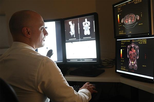 kind of picture does an MRI produce?
An MRI can create exceptionally detailed images of the body’s soft tissues. They can be viewed from all angles, and viewed in cross sections to allow your physician to diagnose illness or injury. Viewing the focus area from multiple vantage points makes a diagnosis more accurate and can help create a treatment plan customized to a specific patient’s needs.
The machine uses strong magnetic fields and radio waves to capture detailed black and white images on a computer, which a physician then reviews. Physicians may use contrast dye or a high contrast scanner to make certain areas stand out more clearly, like in the image.
True Open MRIs for Children
Sometimes, it can be difficult for small children to sit still long enough to achieve a quality image during an MRI. This is even more difficult if the child or parent has anxiety about the procedure, noise, or being alone.
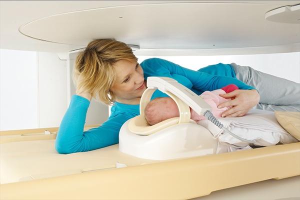 A True Open MRI can help to ease that anxiety, and make the process a little bit easier for everyone involved. Here are just a few ways that a True Open MRI can make the scan easier for both parent and child:
- A parent can stay in the room and even hold/comfort their child throughout the exam if need be.
- An MRI that is open on all four sides can be much less scary for a child, as they can see around the room the whole time.
- A True Open MRI allows space for a child to bring a favorite blanket or stuffed animal with them during the exam, which can serve as a source of comfort.
- For very small children, a parent can be positioned on the True Open MRI bed to hold and protect the child.
When it comes to diagnostic procedures involving children, a caring and compassionate medical staff is everything. Choosing a radiology facility that has ample experience working with children, and treats them with the utmost kindness and understanding, is crucial to a stress-free visit.
“The technologists were amazing with my son! Will always go here for any imaging needs!” -Mother of a Medical Imaging of Fredericksburg patient
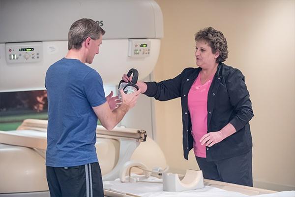
What is it like to get a True Open MRI?
How to prepare for a True Open MRI
Before the exam:
- T
 ell your doctor about any concerns or issues you may have, including metal or electronic devices in your body, like a pacemaker.
 - Avoid excess alcohol and caffeinated beverages.
- Wear comfortable clothes, and prepare to change into a hospital gown if needed.
- Avoid wearing any jewelry or metal.
- Talk to your doctor about any restrictions on eating and/or drinking before the exam.
- Feel free to create your own relaxing playlist to listen to during the exam.
During the exam:
- Your doctor may inject or have you ingest contrast fluid to assist with the clarity of the images.
- You will lay down on a moveable bed and get comfortable before being moved into the machine. A soft antenna device, called a coil, will be placed over or around the area of the body to be imaged.
- Relax, close your eyes, and remain as still as possible.
- You will hear a thumping sound during the exam, which can be muffled by headphones or the music of your choice.
- An MRI usually takes between 20-50 minutes to complete.
- After an MRI, you are free to go about your daily activities as normal.
- Be sure to drink plenty of water to help flush the contrast fluid from your system.

What did we do before the MRI?
Before the widespread use of MRI machines, which began to gain ground in the 1980s, X-rays and, eventually, CT scans, were the only way to non-surgically view the body’s internal structures.
While X-rays and CT scans are still important diagnostic tools, X-rays are two dimensional, making it difficult to see around or between areas. X-rays also don’t differentiate between soft tissue of similar densities, making it difficult to pinpoint specific issues. The use of radiation also limits the amount of people who are qualified to have an X-ray performed and the number of times an individual should have an X-ray scan in a specific time period.
By compiling many X-rays, a CT scan can provide more of a three-dimensional view, but it still uses the same technology, and as such is limited in its differentiation abilities. The use of radiation also limits the scan in the same way as a traditional X-ray.
CT scans and X-rays work well when a physician needs to quickly look at injuries or abnormalities. They are also used if a patient has too much metal in their body for an MRI, or is unable to have an MRI. CT scans and X-rays remain a cost effective diagnostic tool.
When was the MRI invented?
The invention of the MRI is attributed to scientist and physician Raymond Damadian in 1977. Damadian discovered healthy tissue and cancerous tissue reacted to magnetic resonance in different ways, as the amount of water present in each type of tissue fluctuated (tumors contain a higher percentage of water). Similar to the modern MRI, Damadian hypothesized that protons in cancerous tissue would take longer to return to a stable magnetic field than protons in healthy tissue, and thus physicians could locate and isolate problem areas.
MRIs use a rotating magnetic field (discovered in 1882 by Nikola Tesla). Because of this discovery, all MRIs use Tesla units as terms of measurement for the magnetic field.


