
You now have access to high-quality, low-cost ultrasound in King George at our newest outpatient facility. Located at 11131 Journal Pkwy, Medical Imaging of King George offers ultrasound imaging services by appointment Monday through Friday from 8:30 a.m. to 5:00 p.m.
An ultrasound is a relatively simple procedure, but it’s one of the most important steps in identifying the care you need. Your doctor may have ordered this test to examine your organs or diagnose a condition.
With Medical Imaging of King George opening in the community, you’ll have Board Certified, Fellowship Trained Radiologists ready to serve you, without having to drive all the way to Fredericksburg or step foot in a hospital.
- Less wait time
- Highly skilled technologist will administer the ultrasound
- Board Certified, Fellowship Trained Radiologist
- X-ray also available
We are conveniently located by King George Pediatrics. We are near King George Elementary School and King George Family YMCA, just up the road from Shiloh Park. To schedule your ultrasound in King George, call 540-741-9729 (XRAY).
Why Choose Ultrasounds at Medical Imaging of King George?
Getting your ultrasound done by true experts can make an impact on your care. It’s crucial to find a radiology practice with Board Certified Radiologists who are Fellowship Trained in the particular subspecialty you need. When a highly-skilled specialist interprets your images, like the doctors at Medical Imaging of King George, you’ll have greater peace of mind knowing that your results are accurate.
Radiologists at Medical Imaging of King George are accessible and available to your doctor to provide insights to manage your care.
In partnership with Radiologic Associates of Fredericksburg and Mary Washington Healthcare, we offer access to experts from one of the top 80 radiology groups in the nation. Our Board Certified, Fellowship Trained Radiologists serve the community in five counties in Virginia with convenient access to the highest level of care for the greatest value in the region.
As the only medical imaging group in the Commonwealth of Virginia to receive the Diagnostic Imaging Center of Excellence (DICOE) designation, Medical Imaging of Fredericksburg is the largest provider of radiology and vascular surgery services in the area. Medical Imaging of King George is the newest of six locations, including Medical Imaging of Fredericksburg on Sam Perry Boulevard, Medical Imaging of North Stafford, the Imaging Center for Women on Hospital Drive, Medical Imaging at Lee’s Hill, and the Imaging Center for Women at North Stafford.
What is an ultrasound?
Ultrasound, otherwise known as ultrasound scanning or sonography, is one of the most common forms of medical imaging.
An ultrasound machine uses high-frequency sound waves to take pictures of the inside of the body. These aren’t just regular sound waves that we can hear. Ultrasound uses high-frequency sound pulses that go into the body to capture images that help diagnose a variety of conditions or injuries. The images taken during an ultrasound examination can be recorded digitally.
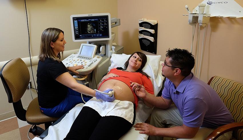
You may be familiar with “obstetrical” ultrasound, commonly used on pregnant women to monitor and track the growth of babies. There are many different types of ultrasound examinations like vascular, abdominal, thyroid and liver—just to name a few.
Ultrasound exams are:
- Quick
- With minimal discomfort
- Radiation-free
- Non-invasive
Top 5 common ultrasound terms:
- Ultrasound imaging– The use of high-frequency sound waves to take images of the body. Also known as ultrasound scanning or sonography.
- Sonogram– Sonogram refers to the image produced by an ultrasound examination.
- Sonographer– A licensed technician who is trained to conduct ultrasound exams.
- Transducer– A small device connected to the ultrasound machine. A sonographer uses a transducer to take the images prescribed by your referring physician.
- Radiologist– A medical doctor who is trained in reading and interpreting ultrasound images.
How does an ultrasound work?
Ultrasound technology is amazing. Using nothing but sound waves, detailed images of the body’s structures and tissues are created—all in a completely non-invasive manner. It’s safe, painless, and radiation-free. So how does it actually work?
According to the Radiological Society of North America (RSNA), “Ultrasound imaging is based on the same principles involved in the sonar used by bats, ships, and fishermen. When a sound wave strikes an object, it bounces back, or echoes. By measuring these echo waves, it is possible to determine how far away the object is as well as the object’s size, shape, and consistency (whether the object is solid or filled with fluid). In medicine, ultrasound is used to detect changes in appearance, size or contour of organs, tissues, and vessels or to detect abnormal masses, such as tumors.”
Ultrasound imaging uses a small transducer (probe) to send high-frequency sound waves into the body. The probe collects the sounds that bounce back, and then a computer uses those sound waves to create an image. Unlike X-rays, ultrasound examinations do not use radiation, thus there is no radiation exposure to the patient. Because ultrasound images are captured in real-time, they can show the structure and movement of the body’s internal organs, blood flowing through blood vessels, or the movement and heartbeat of an unborn baby.
Why do I need an ultrasound?
There are many symptoms that may indicate a need for an ultrasound. If you suffer from one or more symptoms, your doctor may provide you with a referral to an imaging facility. Ultrasounds are a useful tool in examining internal vessels and organs. The images produced during an examination can help diagnose a variety of conditions and diseases.
Top four signs you need an ultrasound:
- Pain
- Swelling
- Infection
- Lumps
What are the different types of ultrasound exams?
There are several different types of ultrasound examinations. This form of medical imaging is frequently used to create images of a variety of different organs.
The following internal organs can be examined via ultrasound:
- Heart
- Blood vessels
- Liver
- Gallbladder
- Spleen
- Pancreas
- Kidneys
- Bladder
- Uterus
- Ovaries
- Developing fetal organs
- Eyes
- Thyroid and parathyroid glands
- Scrotum (testicles)
Ultrasound examinations include:
- Vascular
- Abdominal Renal
- Obstetrical
- Pelvic
- Breast
- Thyroid
- Scrotal
- Musculoskeletal
- Liver Elastography
- AAA and Carotid Wellness Screening
What is carotid wellness screening or carotid ultrasound?
A carotid wellness screening is a painless, non-invasive, non-radioactive test that can determine if your carotid arteries are narrowed. Your carotid arteries run on either side of the neck. Narrowed carotid arteries can increase your risk of stroke. These arteries can become narrow from fat, cholesterol, calcium, or other substances.
Should I have a carotid ultrasound?
Your doctor may recommend a carotid ultrasound if you have health problems that could put you at high risk for a stroke, such as:
- High blood pressure
- Diabetes
- High cholesterol
- Family history of stroke or heart disease
- Recent transient ischemic attack (TIA) or stroke
- Abnormal sound in carotid arteries detected by your doctor using a stethoscope
- Coronary artery disease
How should I prepare for a carotid wellness screening?
A carotid wellness screening is a very simple, quick scan of the arteries in the neck. You will not need to do anything special to prepare for a carotid ultrasound, except for maybe not wearing a shirt that is tight against the neck or dangling earrings. The scan is completely painless and does not use radiation.
What should I expect during a carotid wellness screening?
During a carotid ultrasound, your technician will most likely have you lie down, and apply warm gel to the area around your carotid arteries. Then, they will use a small device called a transducer that emits sound waves to gently examine the arteries. You shouldn’t feel any pain or discomfort during the procedure. You should be able to resume your normal activities right away.
After my screening, how can I reduce my risk of stroke?
Harvard Medical School states that “You can do more than you think to avoid a fatal or debilitating ‘brain attack.’ Strokes don’t usually come out of the blue. Some things you can’t do much about, like age and family history of stroke. But even when an underlying medical condition puts you at risk, you might be able to do something about it. Stroke is potentially one of the most devastating illnesses that we see, and it’s especially tragic when simply taking good care of one’s blood pressure or some other preventive measure might have averted it,” says Thomas Lee, MD, co-editor in chief of the Harvard Heart Letter.
Some very simple ways to reduce your risk of stroke include:
- Losing weight
- Getting your blood pressure under control
- Exercise regularly
- Reduce your alcohol consumption
- Reduce your consumption of salt (sodium)
- Eat a healthy, plant-based diet
What is a Doppler ultrasound?
Doppler ultrasound refers to the technique used to examine blood flow. While ultrasound uses high-frequency sound waves to generate images of the body, a doppler ultrasound specifically bounces soundwaves off red blood cells to measure the flow of blood through the body.
According to the RSNA, “Doppler ultrasound, a special application of ultrasound, measures the direction and speed of blood cells as they move through vessels. The movement of blood cells causes a change in pitch of the reflected sound waves (called the Doppler effect). A computer collects and processes the sounds and creates graphs or color pictures that represent the flow of blood through the blood vessels.”
Why are Doppler ultrasound images important?
Doppler ultrasound images are important because they can help diagnose a variety of conditions. Your doctor may recommend a Doppler ultrasound to identify a procedure or care plan to address problems with your health.
A Doppler ultrasound can detect the following:
- blockages to blood flow, i.e., clots
- narrowing of vessels
- tumors and congenital vascular malformations
- restricted blood flow to organs
- increased blood flow
3 different types of Doppler ultrasound:
- Color Doppler through the use of a computer, measurements are converted into colors to highlight the speed and direction of blood flow.
- Power Doppler provides more detail regarding blood flow than a color Doppler; however, it does not reveal the direction of blood flow.
- Spectral Doppler provides a graphic measurement of blood flow. This information can also be converted into a spectrum of sound with each heartbeat.
What can I expect during my ultrasound exam?
You might experience a little discomfort if the area is tender; but generally, ultrasounds are non-invasive and quick.
During your ultrasound, you may be asked to lie down on an examination table. A medical ultrasound gel will be applied to the area being examined to create a bond between your skin and the ultrasound transducer. Your sonographer will gently press the transducer along the area to capture the images you need for your diagnosis. A special computer will capture these pictures and record measurements.
How should I prepare for my ultrasound?
Most ultrasounds do not require any preparation. When you call to schedule your exam, you may be given some instructions. For example:
- Certain exams require fasting from eating or drinking for up to six hours before your appointment.
- Ultrasounds of the pelvic area may require a full bladder. Depending on your age, you will be asked to drink 8 to 32 ounces of fluids an hour before your appointment.
- You may be asked to remove jewelry or to change into a gown.
In general, you can resume your normal activities as soon as your exam is complete.
What if the ultrasound is for my child?
If your child needs to be scheduled for an ultrasound for the first time, they could be feeling somewhat nervous. Being shy of doctors is normal and understandable!
Advise your child that an ultrasound machine is a special tool that’s like a microphone and camera all-in-one and that this tool helps doctors see inside the body so that they can help keep us healthy. The best part is that your child will get a chance to see the pictures on the computer screen as they are being taken.
You can reassure your child that they are not in any danger, and ask your sonographer to test the transducer on you first, so your child can see that they have nothing to fear.
Make an Appointment Today.
For your convenience, we’re located by King George Pediatrics at 11131 Journal Pkwy, King George, VA 22485.
For both ultrasound and mammography, our hours of operation are Monday through Friday from 8:30 a.m. to 5:00 p.m.
To schedule your ultrasound in King George, call 540-741-9729 (XRAY). We look forward to serving your ultrasound needs.


